Bone Cross Section Diagram - Skeletal System Labeled Diagrams Of The Human Skeleton : Bone marrow cross section stock illustrations.. Medically reviewed by the healthline medical network — written by the healthline editorial team — updated on january 20, 2018. Smartdraw includes 1000s of professional healthcare and anatomy chart templates that you can modify and make your own. The first illustrates the anatomy of a window and frame. Related posts of cross section of a long bone bone test anatomy and physiology. The star of the show (brain) is easily recognizable because it appears highly convoluted, full of ridges (gyri) and indentations (sulci).the paired thalami appear as two circular masses in the midline, forming the walls of the third ventricle.the neurocranium appears as a meshwork (trabecular.
Shop bone cross section diagram label created by chartsanddiagrams. For example, to read this diagram literally, since the cartilage can be seen inside the cutaway section of bone, it incorrectly indicates that the cartilage in fact goes through the bone structure, rather than just being found around the bone end. Bone cross section diagram chart bone bones bone diagram bone chart anatomy biology science. (b) in this micrograph of the osteon, you can clearly see the concentric lamellae and central canals. (b) in this micrograph of the osteon, you can clearly see the concentric lamellae and central canals.
Cross Section Bone High Resolution Stock Photography And Images Alamy from c8.alamy.com Skeletal muscle histology description with labeled diagram well, let's me describe the skeletal muscle histology with labeled diagram so that you might understand every single structure. That's here these 2 diagrams come into play. Bone test anatomy and physiology 12 photos of the bone test anatomy and physiology anatomy and physiology bone lab test, anatomy and physiology bone markings test, anatomy and physiology bone practical test, anatomy and physiology bone tissue test, anatomy and physiology test on bone tissue, bone, anatomy and. I don't like way you've shown the cartilage. So, this is the cross section of skeletal muscle (skeletal muscle of tongue). Related posts of cross section of human bone diagram bone in arm pictures. (b) in this micrograph of the osteon, you can clearly see the concentric lamellae and central canals. The first illustrates the anatomy of a window and frame.
Cross section of long bone diagram | quizlet.
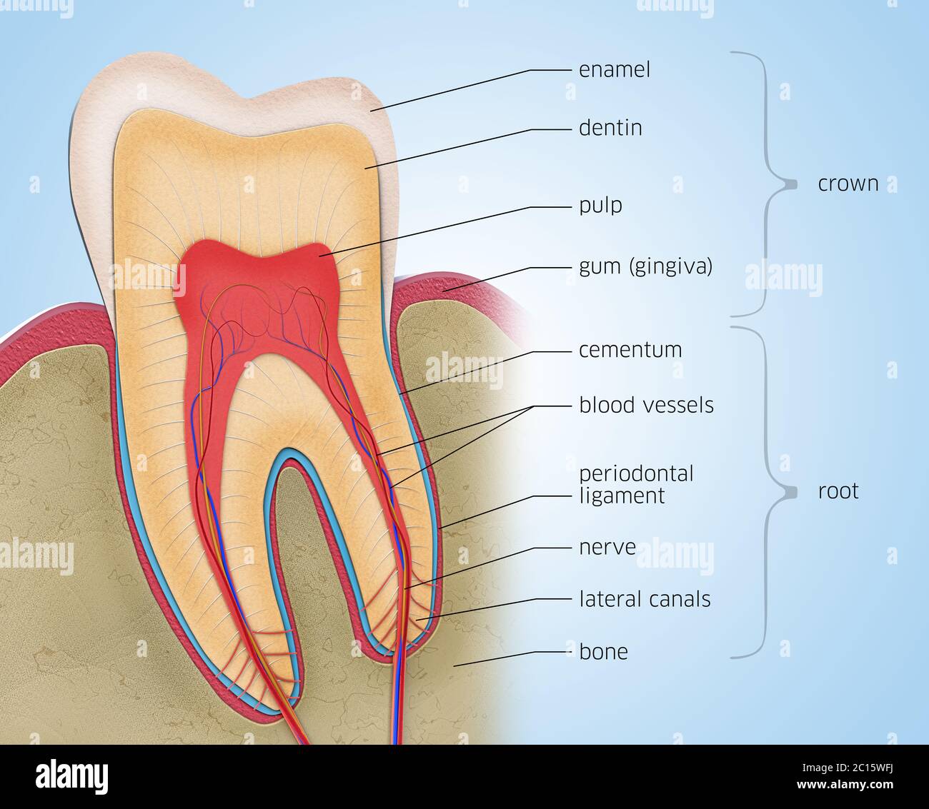
Über 7 millionen englischsprachige bücher. Cross section through the thalamus: It seems confusing and misleading. Cross section of long bone diagram | quizlet. When you're replacing windows, it's nice to know all the different parts of the window and window frame. Skeletal muscle histology description with labeled diagram well, let's me describe the skeletal muscle histology with labeled diagram so that you might understand every single structure. Contact your company to license this image. Personalize it with photos & text or purchase as is! This diagram depicts anatomy of the human eye cross section view.human anatomy diagrams show internal organs, cells, systems, conditions, symptoms and sickness information and/or tips for healthy living. Vector illustration with detailed information isolated on a white background. Human bone, cross section diagram of femur showing osteon, veins, marrow. So, this is the cross section of skeletal muscle (skeletal muscle of tongue). Diagram with articular cartilage, marrow, medullary cavity and periosteum.
Über 7 millionen englischsprachige bücher. Personalize it with photos & text or purchase as is! Cross section of bone diagram. When you're replacing windows, it's nice to know all the different parts of the window and window frame. The star of the show (brain) is easily recognizable because it appears highly convoluted, full of ridges (gyri) and indentations (sulci).the paired thalami appear as two circular masses in the midline, forming the walls of the third ventricle.the neurocranium appears as a meshwork (trabecular.
Cross Section Bone Human High Resolution Stock Photography And Images Alamy from c8.alamy.com Cross section of long bone diagram | quizlet. Download 706 bone cross medical section stock illustrations, vectors & clipart for free or amazingly low rates! Human health concept useful for medical, anatomy and biology educational poster design. (b) in this micrograph of the osteon, you can see the concentric lamellae around the central canals. Related posts of cross section of a long bone bone test anatomy and physiology. The first illustrates the anatomy of a window and frame. It seems confusing and misleading. That's here these 2 diagrams come into play.
For example, to read this diagram literally, since the cartilage can be seen inside the cutaway section of bone, it incorrectly indicates that the cartilage in fact goes through the bone structure, rather than just being found around the bone end.
Contact your company to license this image. (b) in this micrograph of the osteon, you can clearly see the concentric lamellae and central canals. Cross section of bone diagram. New users enjoy 60% off. So, this is the cross section of skeletal muscle (skeletal muscle of tongue). Diagram orienting yourself within such a cross section is easy. Cross section through the thalamus: The star of the show (brain) is easily recognizable because it appears highly convoluted, full of ridges (gyri) and indentations (sulci).the paired thalami appear as two circular masses in the midline, forming the walls of the third ventricle.the neurocranium appears as a meshwork (trabecular. Related posts of cross section of human bone diagram bone in arm pictures. It seems confusing and misleading. Über 7 millionen englischsprachige bücher. I don't like way you've shown the cartilage. Related posts of cross section of a long bone bone test anatomy and physiology.
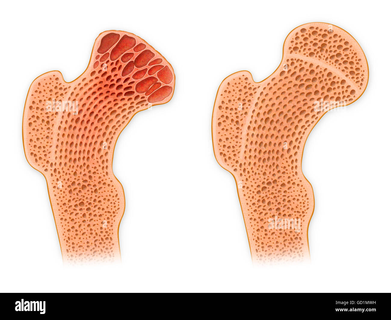
The first illustrates the anatomy of a window and frame. I don't like way you've shown the cartilage. Bone in arm pictures 12 photos of the bone in arm pictures bone cancer arm pictures, pictures of bone cancer in arm, bone, bone cancer arm pictures, pictures of bone cancer in arm. Bone marrow is the soft, highly vascular and flexible connective tissue within bone cavities which serve as the primary site of new blood cell production or bone marrow is the primary source of pluripotent stem cells that give rise to all hemopoietic cells (blood cells) including lymphocytes. For example, to read this diagram literally, since the cartilage can be seen inside the cutaway section of bone, it incorrectly indicates that the cartilage in fact goes through the bone structure, rather than just being found around the bone end.
Diagram Cross Section Of Bone Diagram Quizlet from o.quizlet.com (b) in this micrograph of the osteon, you can clearly see the concentric lamellae and central canals. Bone cross section diagram chart bone bones bone diagram bone chart anatomy biology science. Smartdraw includes 1000s of professional healthcare and anatomy chart templates that you can modify and make your own. Human health concept useful for medical, anatomy and biology educational poster design. Femur bone structure femur bone structure. Personalize it with photos & text or purchase as is! It seems confusing and misleading. Cross section of bone diagram.
The star of the show (brain) is easily recognizable because it appears highly convoluted, full of ridges (gyri) and indentations (sulci).the paired thalami appear as two circular masses in the midline, forming the walls of the third ventricle.the neurocranium appears as a meshwork (trabecular.
Contact your company to license this image. Bone marrow cross section stock illustrations. The first illustrates the anatomy of a window and frame. The star of the show (brain) is easily recognizable because it appears highly convoluted, full of ridges (gyri) and indentations (sulci).the paired thalami appear as two circular masses in the midline, forming the walls of the third ventricle.the neurocranium appears as a meshwork (trabecular. The bladder, like the stomach, is an expandable. Browse 4,294 bone cross section stock photos and images available, or search for human bone cross section to find more great stock photos and pictures. For example, to read this diagram literally, since the cartilage can be seen inside the cutaway section of bone, it incorrectly indicates that the cartilage in fact goes through the bone structure, rather than just being found around the bone end. Bone in arm pictures 12 photos of the bone in arm pictures bone cancer arm pictures, pictures of bone cancer in arm, bone, bone cancer arm pictures, pictures of bone cancer in arm. Vector illustration with detailed information isolated on a white background. It seems confusing and misleading. Cross section through the thalamus: (b) in this micrograph of the osteon, you can see the concentric lamellae around the central canals. Human health concept useful for medical, anatomy and biology educational poster design.
The star of the show (brain) is easily recognizable because it appears highly convoluted, full of ridges (gyri) and indentations (sulci)the paired thalami appear as two circular masses in the midline, forming the walls of the third ventriclethe neurocranium appears as a meshwork (trabecular bone cross section. Skeletal muscle histology description with labeled diagram well, let's me describe the skeletal muscle histology with labeled diagram so that you might understand every single structure.


0 Komentar